Technical note: Measuring cell aggregate diameter using ImageJ
- stemcore
- Sep 11, 2024
- 1 min read
Updated: Oct 8, 2024
Contributor: Dr Mahfuz Chowdhury
ImageJ is one of the most widely used image processing and quantifying app in Life science research. This note will describe how to measure stem cell aggregate diameter using ImageJ Fiji version.
STEPS:
Start ImageJ Fifi app and open the image to be processed by drag and drop on the app toolbar.
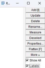
Open ROI Manager by clicking Aanlyze > Tools > ROI Manager and tick "Select All" and "Labels" options
From the app toolbar, select "Straight line" tool.

Draw a line across an aggregate in the image and press "t" key on the keyboard. Follow the same procedures for all other aggregates. The app will automatically number the lines.

Click "Measure" button in the ROI Manager
The app will show various measurements in the "Results" window.

The "Length" variable is the length of the lines in pixels and roughly corresponds to the diameter of the aggregates.
Copy the data in a spreadsheet application for further processing e.g., converting pixel value to metric value by using pixel to metric value conversion ratio from the microscope that was used for the image capture.
Note that this manual procedure is time-consuming for processing many images. Let us know the availability of any automated procedure by comment or email at stem.core@uq.edu.au.


Comments